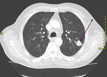
CT scan showing a cancerous tumor in the left lung
Performing a chest radiograph is one of the first investigative steps if a person reports symptoms that may suggest lung cancer. This may reveal an obvious mass, widening of the mediastinum (suggestive of spread to lymph nodes there), atelectasis (collapse), consolidation (pneumonia) or pleural effusion. CT imaging is typically used to provide more information about the type and extent of disease. Bronchoscopy or CT-guided biopsy is often used to sample the tumor for histopathology.
Lung cancer often appears as a solitary pulmonary nodule on a chest radiograph. However, the differential diagnosis is wide. Many other diseases can also give this appearance, including metastatic cancer, hamartomas, and infectious granulomas such as tuberculosis, histoplasmosis and coccidioidomycosis. Lung cancer can also be an incidental finding, as a solitary pulmonary nodule on a chest radiograph or CT scan done for an unrelated reason.The definitive diagnosis of lung cancer is based on histological examination of the suspicious tissue in the context of the clinical and radiological features.
Clinical practice guidelines recommend frequencies for pulmonary nodule surveillance. CT imaging should not be used for longer or more frequently than indicated as extended surveillance exposes people to increased radiation.
 Female Cancer Specialist Doctor in Kanpur female, doctor, cancer, specialist, female, oncologist, oncology, medical, oncologist, chemotherapy, breast, blood, mammography, prostate, center, hospital, radiotherapy, surgery, mouth, kanpur, sn, memorial, female, doctor, cancer, specialist,


